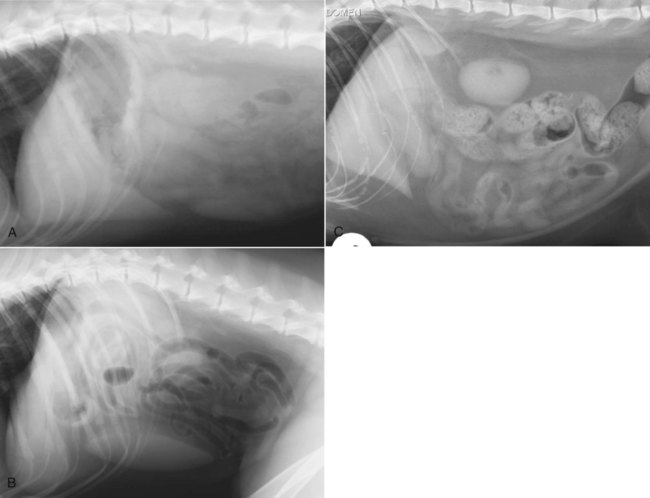
Answered by Liza Treutel on Tue Mar 30 2021 848 PM. In another dog the margins of the liver lobe are rounded and seen caudal to the stomach B.
Architecture Of The Liver And Biliary Tract
If your animal has normal liver enzymes we would expect them to be as shown below.
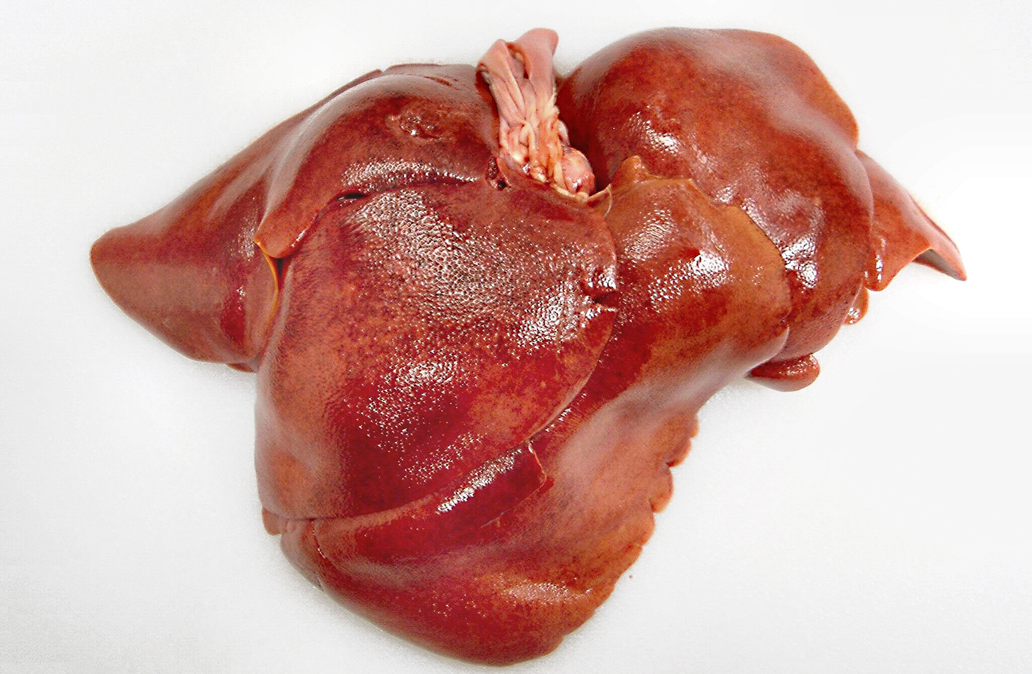
Normal canine liver. Gelatin sponge particles GSPs were injected through a microcatheter for selective embolization of the left hepatic artery in normal dogs. The portal vessels white arrow have hyperechoic walls. The gastric axis is perpendicular to the spine a normal variation for a dog with deep thoracic conformation.
The objective of this study. The Normal Canine Abdominal Exam Exam 3. Hepatocellular death hepatitis and cirrhosis.
While the liver is enormously resilient--it can continue to function even after large portions have been removed--the levels of enzymes present in this critical organ can serve as a gauge of your dogs overall health. Elevated ALT enzymes are usually due to cell damage caused by leakage. The distal extremity of the spleen lies immediately caudal to the liver.
Several pathways in the hepatocyte have been shown to contribute cop-per to the biliary pool including lysosomal exocytosis. Left aspect of the liver on a sagittal view. The normal range for the alanine aminotransferase or ALT enzyme which is liver specific is 10-100 UL according to the Canine Liver Disease Foundation.
C Lateral radiograph of the abdomen of a normal cat. The position of the stomach is one parameter that is commonly used as an indicator of liver size. Healthy Dog Liver Enzyme results.
EXAMINATION OF NORMAL CANINE LIVER VESSELS. Chapter 6 - Morphological classification of parenchymal disorders of the canine and feline liver. Or an even larger dog we can come from behind beneath the legs and palpate like that.
The effect of selective transcatheter arterial embolisation TAE using trisacryl gelatine microspheres TGMs in the normal canine liver was investigated. Up under here and then move dorsally and feel up under. The liver has a homogenous parenchyma and is hypoechoic to the spleen S.
In this dog the liver margins come to a point arrow and are seen ventral near field relative to the stomach A. A dogs liver has many responsibilities. The liver lies entirely within the costal arch appearing small.
The objective of this study was to use the ultrasonographic contrast agent SonoVue to evaluate various transit time indices in the normal canine liver to examine the effect of anesthesia on these parameters and to evaluate the safety of this agent. Its not uncommon for the ALT levels to vary widely from dog to dog and most vets will not be alarmed unless your pets ALT levels are at least 3 times the normal rate on multiple readings. Also a negative vitamin A-storing HSC.
Timothy MWANZA Toru MIYAMOTO Masahiro OKUMURA Mitsuyoshi HAGlO and Toru FUJINAGA. Selective embolisation was achieved by injecting TGMs into the left hepatic artery through a. Typically the range for normal AST is reported between 10 to 40 units per liter and ALT between 7 to 56 units per liter.
Selective embolisation was achieved by injecting TGMs into the left hepatic artery. Liver enzymes are shown on blood tests as a number with a normal range next to them this normal range represents the expected levels seen in the vast majority of cases. This is an indication of increased hepatic volumesize.
The normal range depends on the lab your veterinarian uses but most consider anything up to 130 or so as normal. Normal canine liver stained with desmin antibody. To determine the effects of selective transcatheter arterial embolization TAE in the normal canine liver.
There is individual variation in the size of the normal liver in the dog and the cat making the distinction between a mildly enlarged or mildly small liver from a normal liver highly subjective and relatively inaccurate. Normal histology reversible hepatocytic injury and hepatic amyloidosis. Contrast-enhanced ultrasonography a new imaging modality in veterinary medicine can provide data on tissue perfusion.
The fasted ammonia concentration is 175 mcgdL normal range 050 mcgdL. A final pathway for hepatocyte copper is excretion into the bile. 13 1996 ABSTRACT The aim of this study is to provide a description of the ultrasonographic and angiographic anatomy of the hepatic and portal veins in normal dogs.
The biliary system is the major route of excretion of copper from the body. Select Chapter 7 - Morphological classification of parenchymal disorders of the canine and feline liver. Aspirates from a normal canine left and feline right liver.
Preprandial and postprandial 2-hour SBA are 40 mcmolL normal 08 mcmolL and 102 mcmolL normal 030 mcmolL respectively. Ultrasound of a normal canine liver. Be able to just feel the edge of the liver here.
This finding is normal. A serum biochemistry panel is performed with the results in Table A. Breaking down toxins before they enter the body removing waste products from the blood storing energy and giving blood the ability to clot.
Its not a shock at all to see a dog have an Alk-P of 200 even 400. Majority of nucleated cells are large polygonal epithelial cells hepatocytes in clusters that have finely granular basophilic cytoplasm black arrows. Contrastenhanced ultrasonography a new imaging modality in veterinary medicine can provide data on tissue perfusion.
A A HSC right is positive in the perinuclear cytoplasm weakly extending into a cytoplasmic process. Adult Beagle dogs n 5. The other liver enzymes always seem to be behaving its just this one thats wonky.
Mild elevations are generally considered to be 2-3 times higher than the normal range. Dogs30 The high MT content of normal canine liver is unexplained. The effect of selective transcatheter arterial embolisation TAE using trisacryl gelatine microspheres TGMs in the normal canine liver was investigated.
Top best answers to the question What is normal liver enzyme count in dogs. AST A slight increase in the enzyme AST can be indicative of very serious liver problems like cirrhosis and cancer. Smaller dog come with one hand palpate like this.

The Liver Part 1 Normal Appearance Imv Imaging

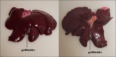
Liver And Spleen Normal Canine Annotated Sonosite M Turbo C11 Imv Imaging

Liver Histology A Normal Liver Dog 1 Portal Area And Periportal Download Scientific Diagram
Testing For Liver Disease Vca Animal Hospital
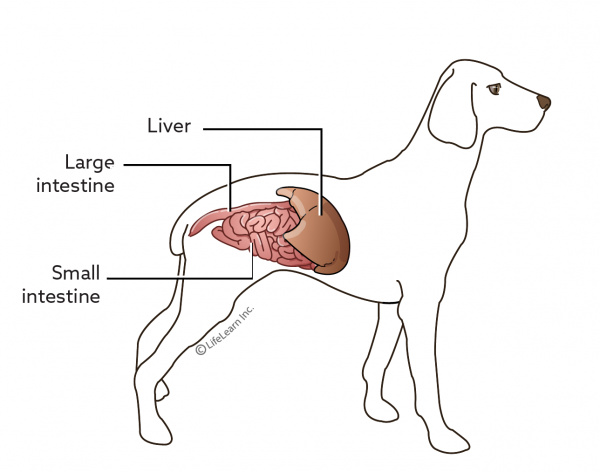
The Liver And Spleen Veterian Key

Liver Dog Gross Appearance Of Liver Showing Pallor Hepatomegaly And Download Scientific Diagram

Animal Liver Disease Long Beach Animal Hospital
Liver Disease In Dogs True Carnivores

Liver Pathology Pictures Flashcards Quizlet
Liver Disease In Dogs Acute Canine Liver Failure The Vet Is In